Thank you for your interest in our ultrasound solutions!
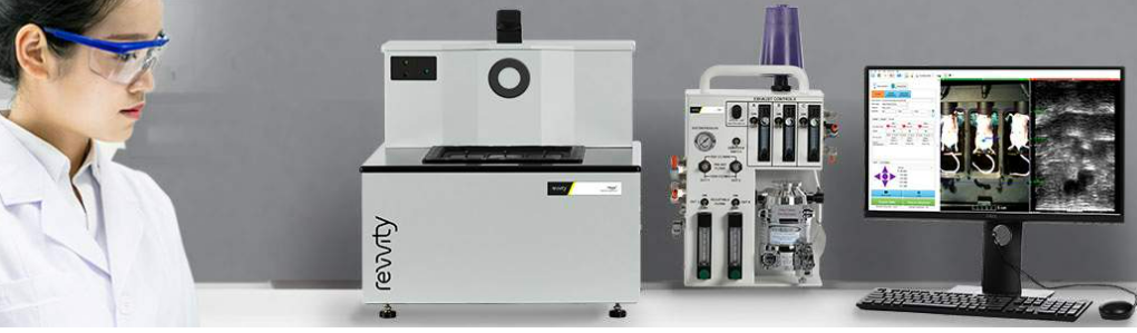
SonoVol is now part of revvity and you can find all the information you need about our products on the revvity website. Click on the links below and you will be redirected to the appropriate product pages.